42 ear anatomy without labels
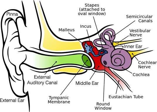
The Ear: Anatomy, Function, and Treatment - Verywell Health The middle ear (also known as the tympanum or tympanic cavity) is a complicated network of tunnels, chambers, openings, and canals mostly inside openings within the temporal bone on each side of the skull. The 2 largest chambers are called the middle ear space and mastoid. Anatomy coloring books: How to use & free PDF | Kenhub Sep 14, 2022 · Generally, an anatomy coloring book will divide subject matter into sections, with each section containing many topics. For each topic you will find black and white anatomical drawings, often accompanied by labels, related text and terminology. Tired of keeping track of so many study materials?
Outer Ear: Anatomy, Location, and Function - Verywell Health Fossa, superior crus, inferior crus, and antihelix: These sections make up the middle ridges and depressions of the outer ear. The superior crus is the first ridge that emerges moving in from the helix. The inferior crus is an extension of the superior crus, branching off toward the head. The antihelix is the lowest extension of this ridge.
Ear anatomy without labels
14,026 Human ear anatomy Images, Stock Photos & Vectors - Shutterstock 14,026 human ear anatomy stock photos, vectors, and illustrations are available royalty-free. See human ear anatomy stock video clips Image type Orientation Color People Artists Sort by Popular Healthcare and Medical Anatomy ear cochlea medicine inner ear organ human body biology diagram Next of 141 heart diagram without labels Blank Ear Diagram | Human Ear Diagram, Ear Anatomy, Ear Diagram . ear diagram blank anatomy human eye drawing unlabeled worksheet parts label ears system quiz senses worksheets special physiology biology college. The Heart - Labelled Diagram wordwall.net. labelled ks4 gcse. DeLand Smiles: February 2012 delandsmiles.blogspot.com Human Body Parts Images Without Labels - Free Vector Download 2020 Human ear diagram with labels and label of anatomy labeling the ear purposegames nose diagram with label diagrams all labels human ear the ear diagram without labels anatomy human charts. Illustration Of Body Parts Labels It is certainly the most widely studied structure the world over. Human body parts images without labels. Download body ...
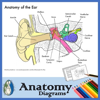
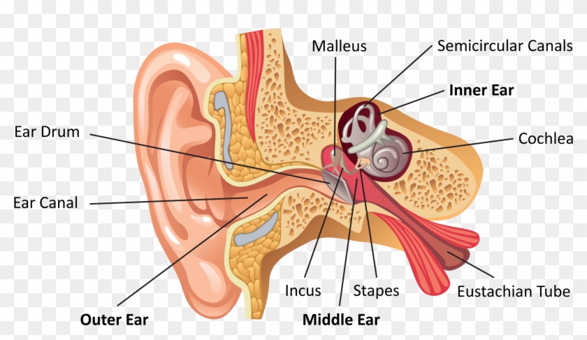
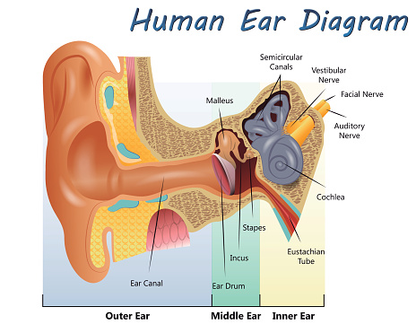
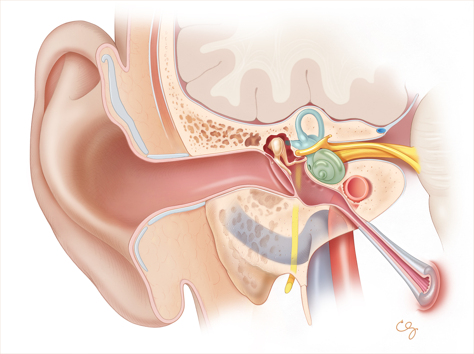
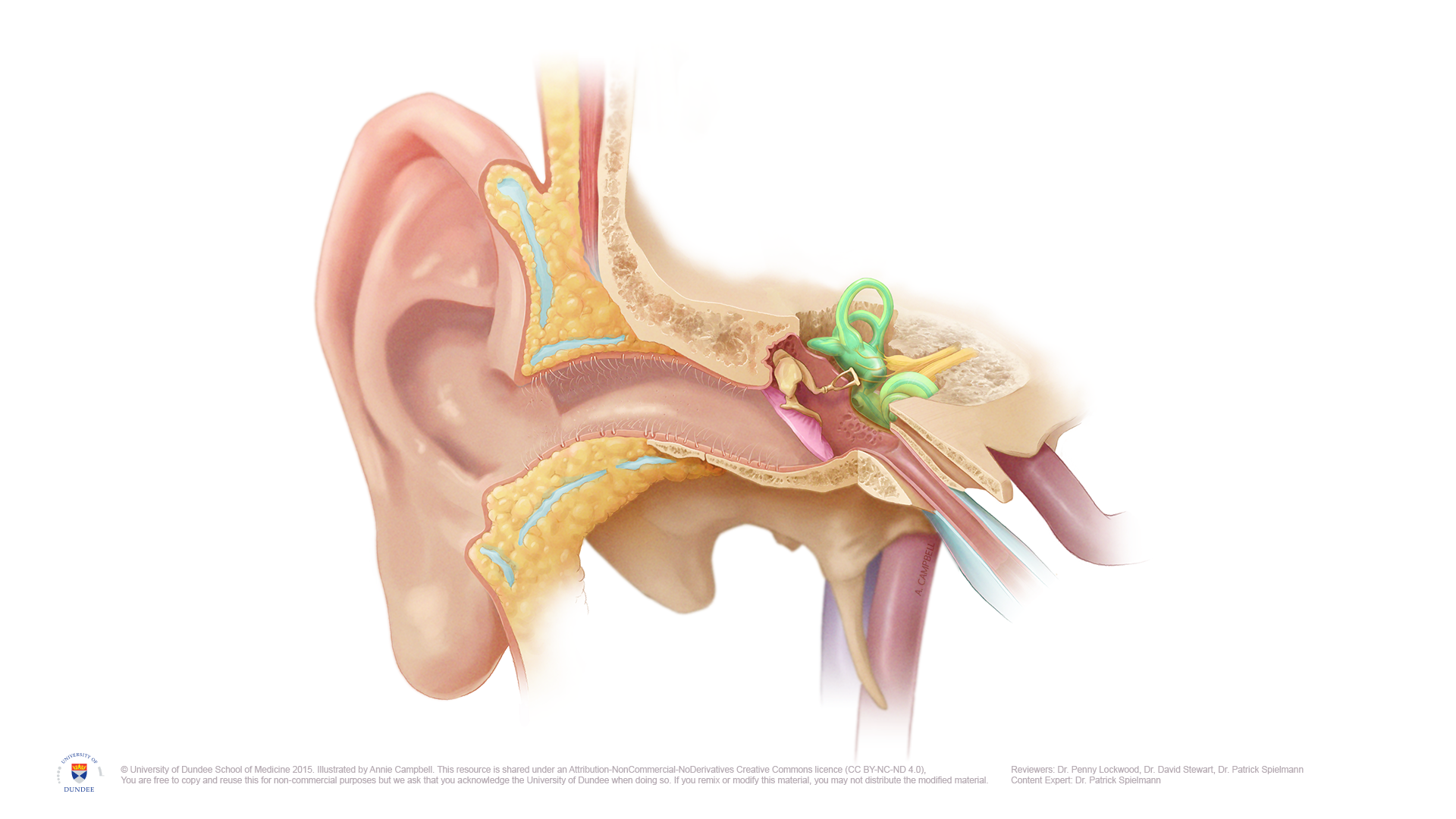
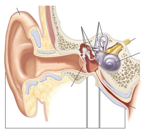
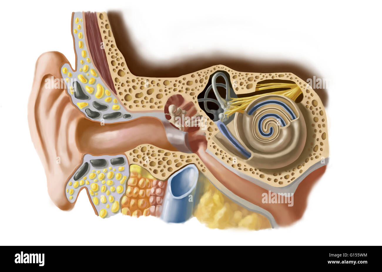
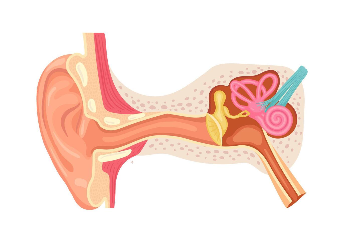
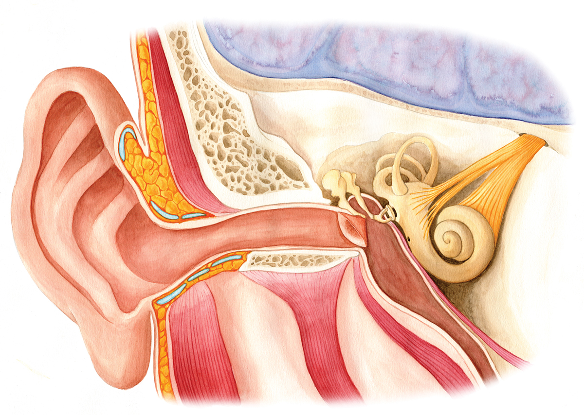
Ear anatomy without labels. Anatomy of the Ear | Inner Ear | Middle Ear | Outer Ear The Outer Ear The outer ear includes: auricle (cartilage covered by skin placed on opposite sides of the head) auditory canal (also called the ear canal) eardrum outer layer (also called the tympanic membrane) The outer part of the ear collects sound. Sound travels through the auricle and the auditory canal, a short tube that ends at the eardrum. Object recognition in medical images via anatomy-guided deep ... Methods. The AAR-DL approach consists of 4 key modules wherein prior knowledge (NI) is made use of judiciously at every stage. In the first module AAR-R, objects are recognized based on a previously created fuzzy anatomy model of the body region with all its organs following the automatic anatomy recognition (AAR) approach wherein high-level human anatomic knowledge is precisely codified. Label Anatomy Dogs Teaching Resources | Teachers Pay Teachers 3.0. (1) $2.50. PDF. Label the Parts of a DogThis is a perfect addition to your Chinese New-year Lessons. Printables - 12 parts• Colour Printout - A control chart for independent work• Printout 1- boxes with dashes for cutting and gluing• Printout 2- with the words at the bottom, for writing Other Dog Lessons:2018 Year of the Dog ~ Dog ... Human Ear Diagram - Bodytomy Auditory Ossicles: The three small bones in the middle ear, called malleus, stapes, and incus, are connected. These bones together are called the auditory ossicles, and their purpose is to let the sound that strikes the eardrum, further into the inner ear.
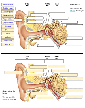
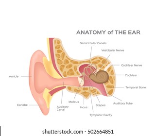
Petrous bone CT: normal anatomy| e-Anatomy - e-Anatomy - IMAIOS Sep 13, 2021 · Anatomy of the temporal bone: how to view the anatomical labels. This module is a comprehensive and affordable learning tool for residents and medical students and specially for neuroradiologists and otolaryngologists. It provides images in the axial and coronal planes, allowing the user to review and learn anatomy interactively. Ear Labels Flashcards | Quizlet Terms in this set (16) auricle external auditory canal tympanic membrane malleus (hammer) Incus (anvil) stapes (stirrup) auditory/eustachian/pharyngotympanic tube vestibules semicicular canals Ampulla of semicircular canals round window oval window and round window cochlea snail cochlear duct in cochlea vestibular nerve Label Parts of the Human Ear - University of Dayton Parts of the Ear. Select the correct label for each part of the ear. Click on the Score button to see how you did. Incorrect answers will be marked in red. Blank ear diagrams and quizzes: The fastest way to learn ear anatomy Ear diagrams (labeled and unlabeled) Accelerate your learning with interactive quizzes Sources + Show all Ear anatomy overview Although it's not obvious to look at, the ear is anatomically divided into three portions: External (outer) ear Middle ear Inner ear As you can imagine, there's a lot of associated anatomy to learn for each portion!
Human Ear: Structure and Anatomy - Online Biology Notes Ear ossicles: The three ear ossicles (malleus, incus and stapes) form a chain of lever extending from tympanic membrane to inner ear. The ear ossicles transmit sound wave from ear drum to inner ear. Ear ossicles communicate the ear drum with internal ear through fenestra ovalis ( oval window). The ear ossicles are; label the ear worksheet 14 Best Images Of Ear Hearing Worksheets - Listening Ear Craft Template ear worksheet diagram inner anatomy answers parts eye animals physiology labelled worksheets ossicles senses auditory middle hearing outer pinna canal Ear Diagram Without Labels & With Them - Labelling Worksheet Image result for ear structure without label | Ear anatomy, Human ear ... Each nephron is made of two main parts called malpighian body and covoluted tubule.Malphigian body is double layered cup also called bowman`s capsule. The inner cup consists network of capillaries called Glomerulus. Now let`s start the diagram. 1.Draw a egg shape and make a folded curve as shown in the figure and… L Amy Yee Anatomy Human Middle Ear Anatomy Cross Section View With Labels Stock Photo ... Description Computer generated image of the human middle ear bones and inner ear with anatomical labeling. 1 credit Essentials collection for this image $4 with a 1-month subscription (10 Essentials images for $40) Continue with purchase View plans and pricing Includes our standard license. Add an extended license. Credit: Hank Grebe
Anatomy of the Ear - Diagrams for Coloring/Labeling, with ...
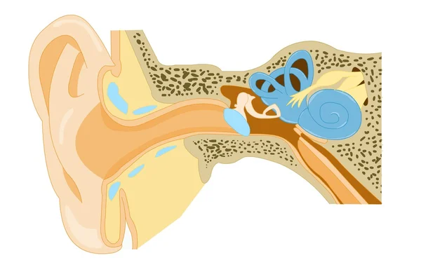
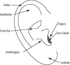
Human Ear Anatomy - Parts of Ear Structure, Diagram and Ear Problems The external (outer) ear consists of the auricle, external auditory canal, and eardrum (Figure 1 and 2). The auricle or pinna is a flap of elastic cartilage shaped like the flared end of a trumpet and covered by skin. The rim of the auricle is the helix; the inferior portion is the lobule. Ligaments and muscles attach the auricle to the head.

Ear Diagram and Labeling Worksheet / Worksheet
Anatomy of the Ear | Geeky Medics Figure 1.Anatomy of the external ear. 4 Innervation of the auricle. The auricle has several sources of sensory innervation:. The superficial surface is supplied by the great auricular nerve and lesser occipital nerve, both of which are branches of the cervical plexus (C2 & C3), and the auriculotemporal branch of the mandibular nerve, which is a branch of the trigeminal nerve (cranial nerve V)

The structure and function of the ear and its role in hearing ...
human ear | Structure, Function, & Parts | Britannica human ear, organ of hearing and equilibrium that detects and analyzes sound by transduction (or the conversion of sound waves into electrochemical impulses) and maintains the sense of balance (equilibrium). The human ear, like that of other mammals, contains sense organs that serve two quite different functions: that of hearing and that of postural equilibrium and coordination of head and eye ...

Ear Anatomy. Illustration Showing the Way of a Sound Wave To ...
FUNDAMENTAL PRINCIPLES OF HUMAN ANATOMY & PHYSIOLOGY Jul 02, 2016 · Labels of human body features displayed on images of actual human bodies, from which body hair and male facial hair has been removed. (Source: City Studios in Stockholm ( ...
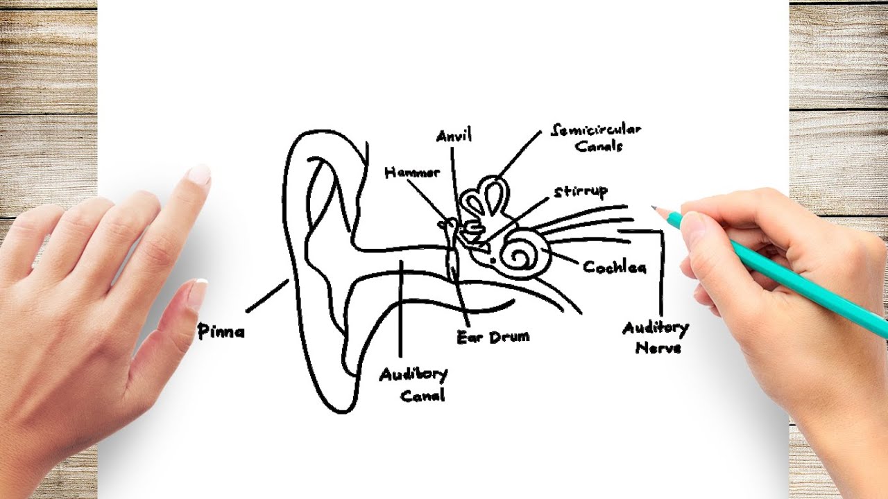
How to Draw Human Ear Diagram With Labelling #HumanEar
Anatomy and Physiology of the Ear - Health Encyclopedia - University of ... The ear is the organ of hearing and balance. The parts of the ear include: Pinna or auricle. This is the outside part of the ear. External auditory canal or tube. This is the tube that connects the outer ear to the inside or middle ear. Tympanic membrane (eardrum). The tympanic membrane divides the external ear from the middle ear.

chapter 10 anatomy labeling the ear Diagram | Quizlet
Best 10 Anatomy Apps - Last Updated July 26, 2022 - AppGrooves May 01, 2018 · This app is absolutely phenomenal. I have used it for 6 years now. The fact that you are able to explore in zoomable detail the thousands of muscles, tendons, ligaments joints, bones, organs, attachment points, connections, surfaces, etc., has been so enlightening for me in exploring the anatomy involved in the random injuries I have passed in life, including injuries that I never knew of ...

The Ear Poster 24x36inch, Anatomy, Organs of Hearing and Balance
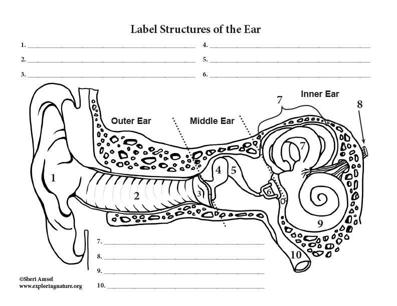
Ear Diagram and Labeling Worksheet / Worksheet - Twinkl This allows you to tailor the task to the individual abilities of your learners. The first worksheet presents an ear with annotations showing the first letters of its key features. For example, a label marked 'P' links to the Pinna (outer ear). The second page shows an ear diagram without labels. The final page shows the labels linking to the ...
External Auditory Canal Of Human Ear (With - Canvas Print | Alan Gesek
Image result for ear structure without label - Pinterest Preschool Kid Learning Ear Coloring Pages to Color, Print and Download for Free along with bunch of favorite Ear coloring page for kids. Simply do online coloring for Preschool Kid Learning Ear Coloring Pages directly from your gadget, support for iPad, android tab or using our web feature. D. Darina Lețu. Doodle and Art.

Label the parts of the ear in the following illustration ...
Normal chest MDCT with anatomic labels | e-Anatomy - IMAIOS Mar 10, 2022 · Normal anatomy of the thorax on labeled Chest CT: radiological anatomy of the lungs, mediastinal lymph nodes, trachea, bronchi, pleural cavity, heart and pulmonary vessels. × Your email address is not verified.

2: Gross anatomy of the human ear including the outer, middle ...
Anatomy, Head and Neck, Ear Ossicles - StatPearls - NCBI Bookshelf The middle ear functions to connect the sound waves from the external environment and transfer them to the inner ear for auditory transduction. The auditory ossicles (malleus, incus, and stapes) play a key role in this function. The malleus connects to the tympanic membrane transferring auditory oscillations to the incus and then the stapes. The stapes connects to the oval window allowing for ...

The ear canal: Anatomy, diagram, and common conditions
Ear (Anatomy): Overview, Parts and Functions | Biology Dictionary The tympanic membrane, or "ear drum" is a thin, tightly-stretched membrane that separates the outer from the middle ear. Just like the membrane of an actual drum, the tympanic membrane vibrates in response to the sounds that are funneled to it by the pinna and ear canal. The outside of the tympanic membrane faces the ear canal.

Cat ear anatomy, illustration - Stock Image - C029/9561 ...
Anatomy of the eye: Quizzes and diagrams | Kenhub How to learn the parts of the eye. Found within two cavities in the skull known as the orbits, the eyes are surrounded by several supporting structures including muscles, vessels, and nerves. There are 7 bones of the orbit, two groups of muscles (intrinsic ocular and extraocular), three layers to the eyeball … and that's just the beginning.

Labelled diagram of the ear
Ear Anatomy - Outer Ear | McGovern Medical School The medical term for the outer ear is the auricle or pinna. The outer ear is made up of cartilage and skin. There are three different parts to the outer ear; the tragus, helix and the lobule. EAR CANAL The ear canal starts at the outer ear and ends at the ear drum. The canal is approximately an inch in length.

Label the ear structures. | Homework.Study.com
Picture of the Ear: Ear Conditions and Treatments - WebMD The ear has external, middle, and inner portions. The outer ear is called the pinna and is made of ridged cartilage covered by skin. Sound funnels through the pinna into the external auditory...
Ear - Anatomy and hearing
Inner Ear Anatomy, Function, and Health Inner ear function. The inner ear has two main functions. It helps you hear and keep your balance. The parts of the inner ear are attached but work separately to do each job. The cochlea works ...
Ear anatomy Stock Photos, Royalty Free Ear anatomy Images ...
Image result for ear structure without label | Ear diagram, Human ear ... Feb 12, 2018 - Image result for ear structure without label. Feb 12, 2018 - Image result for ear structure without label. Pinterest. Today. Explore. When the auto-complete results are available, use the up and down arrows to review and Enter to select. Touch device users can explore by touch or with swipe gestures.

Anatomy of the Inner Ear | Doctor Stock
Cefdinir Antibiotic Side Effects, Uses (Strep, Middle Ear ... Cefdinir is an antibiotic in the cephalosporin drug class prescribed to treat infections, for example, middle ear, tonsillitis, strep throat, bronchitis, and sinusitis. Common side effects are nausea, abdominal pain, loose stools, and vaginitis. Dosage and pregnancy and breastfeeding safety information are included.
Outer Ear Anatomy Neutral Pattern (without center label ...
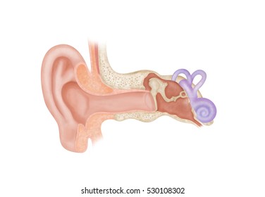
Ear Anatomy without Labels, Digital Art - Shutterstock Ear Anatomy Without Labels Digital Art Stock Illustration 530108302 Download for free See more Popularity score High Usage score High usage Superstar Shutterstock customers love this asset! Item ID: 530108302 Ear Anatomy without Labels, Digital Art Formats 8976 × 6201 pixels • 29.9 × 20.7 in • DPI 300 • JPG
Ear diagram - Teaching resources
Human Body Parts Images Without Labels - Free Vector Download 2020 Human ear diagram with labels and label of anatomy labeling the ear purposegames nose diagram with label diagrams all labels human ear the ear diagram without labels anatomy human charts. Illustration Of Body Parts Labels It is certainly the most widely studied structure the world over. Human body parts images without labels. Download body ...
Ear Anatomy Labeled - Ear Parts Labeled, HD Png Download ...
heart diagram without labels Blank Ear Diagram | Human Ear Diagram, Ear Anatomy, Ear Diagram . ear diagram blank anatomy human eye drawing unlabeled worksheet parts label ears system quiz senses worksheets special physiology biology college. The Heart - Labelled Diagram wordwall.net. labelled ks4 gcse. DeLand Smiles: February 2012 delandsmiles.blogspot.com
Ear Diagram Without Label Vector Free | AI, SVG and EPS
14,026 Human ear anatomy Images, Stock Photos & Vectors - Shutterstock 14,026 human ear anatomy stock photos, vectors, and illustrations are available royalty-free. See human ear anatomy stock video clips Image type Orientation Color People Artists Sort by Popular Healthcare and Medical Anatomy ear cochlea medicine inner ear organ human body biology diagram Next of 141
Anatomy of the inner ear
Ear Anatomy – Outer Ear | McGovern Medical School

Outer ear - Wikipedia

Label Parts of the Human Ear
Dundee - Drawing Anatomy of the ear - No labels | AnatomyTOOL
Auditory pathway: Anatomy, ear structures, transduction | Kenhub
Ear Anatomy Outline Vector Illustration Stock Vector (Royalty ...

Ear Anatomy Labeling Worksheet.docx - Ear Anatomy Labeling ...
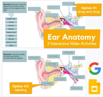
Ear Anatomy - interactive drag-and-drop, labeling in Slides
Label Parts of the Human Auditory System
Parts of the Ear – Fill In the Blank
Ear diagram hi-res stock photography and images - Alamy
Ear Anatomy – Drag and Drop Activity

1: Diagram showing the structure of the human ear, detailing ...
Anatomy of the human ear. Internal structure of the ears ...
Superior Canal Dehiscence | UCI Head and Neck Surgery - UCI ...
Human Ear Structure Medical Educational Science Stock Vector ...
Anatomy of the Ear Parts of the Ear Minimum time needed 12 ...
Ear Anatomy for Visual Guide to ENT Pathology on Behance

Cochlea Implants for the Deaf and Severely Hard of Hearing ...
Ear Anatomy Without Labels Digital Art Stock Illustration ...
Post a Comment for "42 ear anatomy without labels"