40 structure of the heart with labels
Ch. 19 Circulatory System- heart Flashcards | Quizlet Place the labels in order denoting the flow of blood through the pulmonary circuit beginning with the right atrium and ending in the left atrioventricular valve. The first and last structures are given. Right atrium 1. tricuspid valve 2. right ventricle 3. pulmonary valve 4. pulmonary trunk 5. pulmonary artery 6. lungs 7. pulmonary vein Labelling the heart — Science Learning Hub Blood transports oxygen and nutrients to the body. It is also involved in the removal of metabolic wastes. In this interactive, you can label parts of the human heart. Drag and drop the text labels onto the boxes next to the diagram. Selecting or hovering over a box will highlight each area in the diagram.
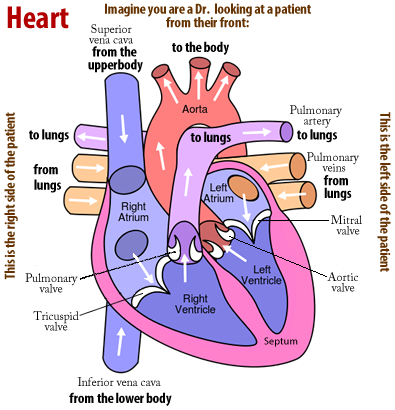
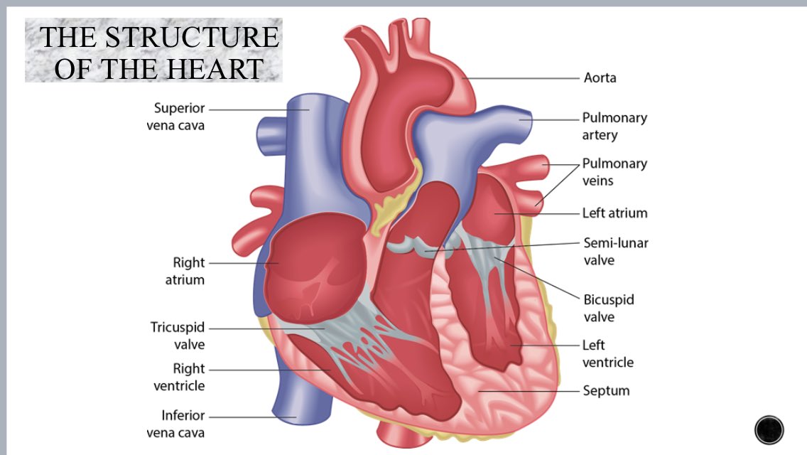
Label the Heart - The Biology Corner A simple heart diagram with arrows and boxes for students to practice labeling the chambers and major vessels. Name:_____Date: _____ Label the Heart. Word Bank: Left Atrium | Right Atrium | Left Ventricle | Right Ventricle Aorta | Pulmonary Veins | Pulmonary Artery | Superior Vena Cava | Inferior Vena Cava Bicuspid Valve | Tricuspid Valve ...
Structure of the heart with labels
Diagrams, quizzes and worksheets of the heart | Kenhub Worksheet showing unlabelled heart diagrams. Using our unlabeled heart diagrams, you can challenge yourself to identify the individual parts of the heart as indicated by the arrows and fill-in-the-blank spaces. This exercise will help you to identify your weak spots, so you'll know which heart structures you need to spend more time studying ... Label Heart Anatomy Diagram Printout - EnchantedLearning.com Label Heart Anatomy Diagram Printout - EnchantedLearning.com The heart is a fist-sized, muscular organ that pumps blood through the body. Oxygen-poor blood enters the right atrium of the heart (via veins called the inferior vena cava and the superior vena cava). The structure of the heart - Structure and function of the heart ... It is located in the middle of the chest and slightly towards the left. The heart is a large muscular pump and is divided into two halves - the right-hand side and the left-hand side. The...
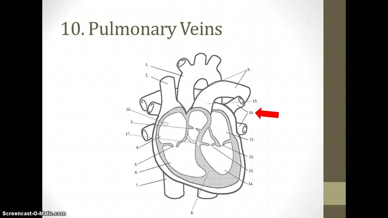
Structure of the heart with labels. Heart Diagram with Labels and Detailed Explanation There are four chambers of the heart. The upper two chambers are the auricles and the lower two are called ventricles. There are four main valves of the human heart- aortic valve, mitral valve, pulmonary valve and tricuspid valve. They help prevent backflow of the blood. The Heart - Science Quiz - Seterra The Heart - Science Quiz Home >> Seterra Anatomy and Science Quizzes >> The Heart The Heart - Science Quiz Aorta, Aortic valve, Left atrium, Left ventricle, Mitral valve, Pulmonary artery, Pulmonary valve, Pulmonary vein, Right atrium, Right ventricle, Septum, Superior vena cava, Tricuspid valve (13) Create custom quiz The Anatomy of the Heart - Quiz 1 - Free Anatomy Quiz The circulatory system - lower body image, with blank labels attached. The circulatory system - a PDF file of the upper and lower body for printing out to use off-line. Describe and explain the function of the circulatory system - The circulatory system consists of the heart, the blood vessels (veins, arteries, and capillaries), and the blood. Heart Labeling Quiz: How Much You Know About Heart Labeling? Here is a Heart labeling quiz for you. The human heart is a vital organ for every human. The more healthy your heart is, the longer the chances you have of surviving, so you better take care of it. Take the following quiz to know how much you know about your heart. Questions and Answers 1. What is #1? 2. What is #2? 3. What is #3? 4. What is #4?
Chapter 19: The Heart Flashcards | Quizlet •Allows heart to beat without friction, gives it room to expand and resists excessive expansion •Parietal pericardium-tough outer, fibrous layer of connective tissue-inner serous layer •Visceral pericardium (a.k.a. epicardium of heart wall)-serous lining of sac turns inward at base of heart to cover the heart surface Heart Diagram – 15+ Free Printable Word, Excel, EPS, PSD ... Teachers and students use the heart diagram, in biological science, to study the structure and functions of a human being’s heart. Friends and colleagues on the other hand may find this diagram template useful when it comes to sending special, personalized gifts to their family members and significant others. Download the template today, and ... 147 Heart Anatomy With Labels Premium High Res Photos Browse 147 heart anatomy with labels stock photos and images available, or start a new search to explore more stock photos and images. heart blood flow - heart anatomy with labels stock illustrations. human heart anatomy. blood flow - heart anatomy with labels stock illustrations. circulatory system, diagram - heart anatomy with labels stock ... The Anatomy of the Heart, Its Structures, and Functions The heart is the organ that helps supply blood and oxygen to all parts of the body. It is divided by a partition (or septum) into two halves. The halves are, in turn, divided into four chambers. The heart is situated within the chest cavity and surrounded by a fluid-filled sac called the pericardium. This amazing muscle produces electrical ...
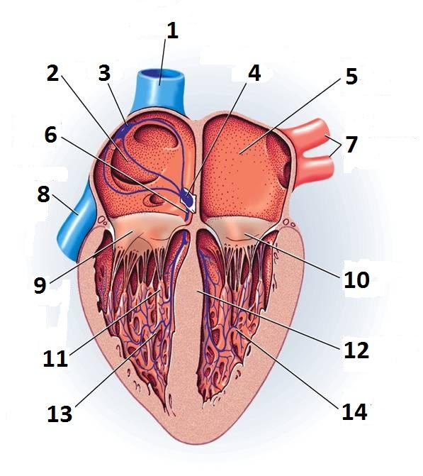
How to Draw the Internal Structure of the Heart (with Pictures) To draw the internal structure of a human heart, follow the steps below. Part 1 Finding a Diagram 1 To find a good diagram, go to Google Images, and type in "The Internal Structure of the Human Heart". Find an image that displays the entire heart, and click on it to enlarge it. 2 Find a piece of paper and something to draw with. Structure of the Heart | SEER Training The human heart is a four-chambered muscular organ, shaped and sized roughly like a man's closed fist with two-thirds of the mass to the left of midline. The heart is enclosed in a pericardial sac that is lined with the parietal layers of a serous membrane. The visceral layer of the serous membrane forms the epicardium. Layers of the Heart Wall Human Heart - Diagram and Anatomy of the Heart - Innerbody The heart is a muscular organ about the size of a closed fist that functions as the body's circulatory pump. It takes in deoxygenated blood through the veins and delivers it to the lungs for oxygenation before pumping it into the various arteries (which provide oxygen and nutrients to body tissues by transporting the blood throughout the body). Label the Heart Diagram | Quizlet Label the Heart STUDY Learn Write Test PLAY Match Created by bluesas9 Terms in this set (15) Superior Vena Cava ... Right Ventricle ... Left Atrium ... Atrioventricular/Tricuspid Valve ... Atrioventricular/Mitral Valve ... Septum ... Right Atrium ... Semi-lunar Valves ... Left Pulmonary Veins ... Right Pulmonary Veins ... Left Pulmonary Arteries
Labeling The Heart - YouTube
Anabolic steroid - Wikipedia Most steroid users are not athletes. In the United States, between 1 million and 3 million people (1% of the population) are thought to have used AAS. Studies in the United States have shown that AAS users tend to be mostly middle-class heterosexual men with a median age of about 25 who are noncompetitive bodybuilders and non-athletes and use the drugs for cosmetic purposes. "

Heart Structures Flashcards | Easy Notecards
Structure Of The Heart | A-Level Biology Revision Notes The heart is a hollow muscular organ that lies in the middle of the chest cavity. It is enclosed in the pericardium, which protects the heart and facilitates its pumping action. The heart is divided into four chambers: The two atria (auricles): these are the upper two chambers. They have thin walls which receive blood from veins.

Heart structure and function — structure and function of the heart cardiac output
Eating for a healthy heart - Baker This may help to improve your overall heart health. Unhealthy ‘saturated and trans’ fats should be limited. This fact sheet will help you with: choosing healthy fats and sources; practical ways to include this into your diet; reducing unhealthy fats in your diet; how to read nutrition food labels. Download fact sheet

Heart Disease: Structure Of The Heart
The 22 Best Luxury Items Our Editors Will Stand By Forever ... Mar 26, 2022 · "As you can see, I'm a big proponent of investing in a designer handbag, and this is the perfect sidekick. The mini bag trend has held a special place in my heart ever since it emerged so I think everyone needs the Le Chiquito bag from Jaquemus. It's the perfect shape and forces me to pack light. Plus, I just love being hands-free."—Mayhew

small intestine histology labeled | Nursing School | Pinterest | Anatomy
Structure of the Heart | The Franklin Institute Structure of the Heart Although most people know that the human heart doesn't bear much resemblance to a heart drawn on a Valentine's Day card, the image can still be a useful way to learn and remember the parts of the heart. The heart consists of four chambers: two atria on the top and two ventricles on the bottom.
34 Label Of Heart Diagram - Labels Database 2020
Human Heart - Anatomy, Functions and Facts about Heart Following are the main functions of the heart: One of the primary functions of the human heart is to pump blood throughout the body. Blood delivers oxygen, hormones, glucose and other components to various parts of the body, including the human heart. The heart also ensures that adequate blood pressure is maintained in the body.
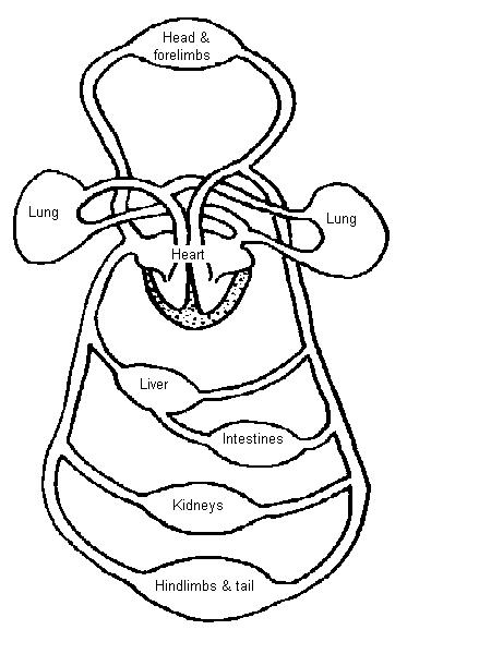
Circulatory System Worksheet - WikiEducator
Layers of the heart: Epicardium, myocardium, endocardium | Kenhub The myocardium is functionally the main constituent of the heart and the thickest layer of all three heart layers. It is a muscle layer that enables heart contractions. Histologically, the myocardium is comprised of cardiomyocytes.Cardiomyocytes have a single nucleus in the center of the cell, which helps to distinguish them from skeletal muscle cells that have multiple nuclei dispersed in the ...
Unlabeled Heart Diagram - Cliparts.co
Heart: Anatomy and Function The parts of your heart are like the parts of a house. Your heart has: Walls. Chambers (rooms). Valves (doors). Blood vessels (plumbing). Electrical conduction system (electricity). Heart walls Your heart walls are the muscles that contract (squeeze) and relax to send blood throughout your body.

PLEASE CAN ANYONE GIVE ME A SYSTEMATIC DIAGRAM OF TH HEART - Science - - 9821775 | Meritnation.com
George B. Moody PhysioNet Challenge | Quick links for this ... Feb 22, 2022 · The public training set contains heart sound recordings, routine demographic information, murmur-related labels (presence, absence, or unknown), outcome-related labels (normal or abnormal), annotations of the murmur characteristics (location, timing, shape, pitch, quality, and grade), and heart sound segmentations.
Limehurst Academy PE on Twitter: "GCSE PE Revision: cardiovascular system structure of heart and ...
BYJUS BYJUS

34 Label Of Heart Diagram - Labels Database 2020
Heart Blood Flow | Simple Anatomy Diagram, Cardiac Circulation ... - EZmed Step 1 and 6 involve a blood vessel, which makes sense as this is how blood enters and exits that side of the heart. Steps 2-5 involve a chamber, valve, chamber, and valve. So if you remember this general pattern, it will help you recall the order in which blood flows through each side of the heart.
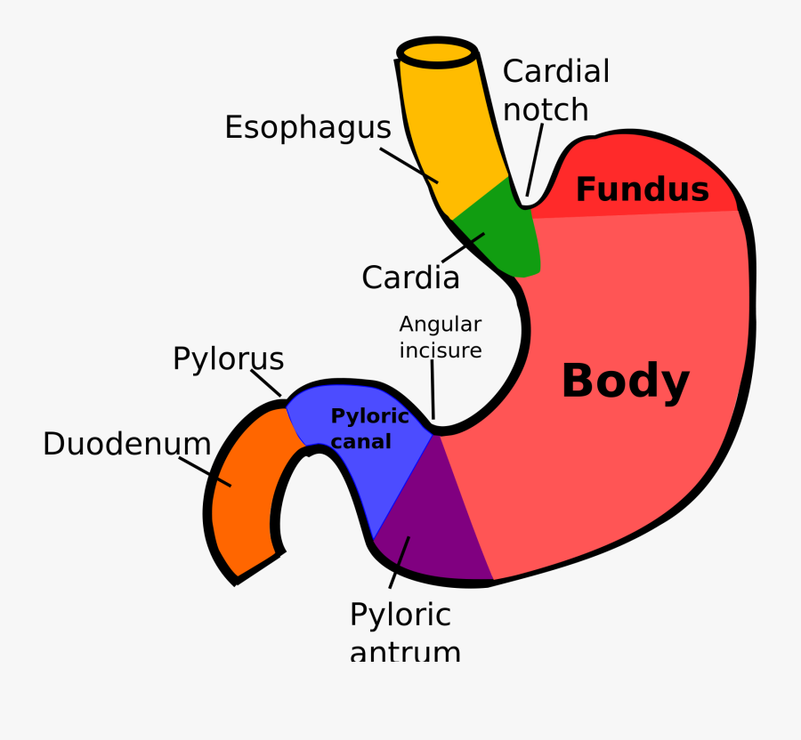
Regions Of Stomach Diagram , Free Transparent Clipart - ClipartKey
Label the heart — Science Learning Hub In this interactive, you can label parts of the human heart. Drag and drop the text labels onto the boxes next to the diagram. Selecting or hovering over a box will highlight each area in the diagram. Pulmonary vein Right atrium Semilunar valve Left ventricle Vena cava Right ventricle Pulmonary artery Aorta Left atrium Download Exercise Tweet
Free Heart Diagram Unlabeled, Download Free Heart Diagram Unlabeled png images, Free ClipArts on ...
Heart Anatomy: Heart Dissection The picture below shows an anterior view of the heart with the pericardium removed. The letters indicated in the text refer to the labels on the picture. The anterior surface of the heart is characterized by the presence of the large arteries leaving the base of the heart, the pulmonary trunk (H) and the aorta (G). The pulmonary trunk is the ...

Flat Style Illustration Of Human Heart Anatomy Stock Illustration - Download Image Now - iStock
Human Heart (Anatomy): Diagram, Function, Chambers, Location in Body The heart is a muscular organ about the size of a fist, located just behind and slightly left of the breastbone. The heart pumps blood through the network of arteries and veins called the...
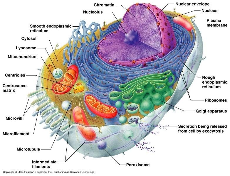
Print Chapter 2 cells flashcards | Easy Notecards
Label the Heart Shows a picture of a heart with letters and blanks for practice with labeling the parts of the heart and tracing the flow of blood within the heart.
Post a Comment for "40 structure of the heart with labels"