40 structure of the heart without labels
File:Diagram of the human heart (no labels).svg - Wikimedia This file is licensed under the Creative Commons Attribution-Share Alike 4.0 International license.: You are free: to share - to copy, distribute and transmit the work; to remix - to adapt the work; Under the following conditions: attribution - You must give appropriate credit, provide a link to the license, and indicate if changes were made. You may do so in any reasonable manner, but ... Structure Of The Heart | A-Level Biology Revision Notes The heart is a hollow muscular organ that lies in the middle of the chest cavity. It is enclosed in the pericardium, which protects the heart and facilitates its pumping action. The heart is divided into four chambers: The two atria (auricles): these are the upper two chambers. They have thin walls which receive blood from veins.
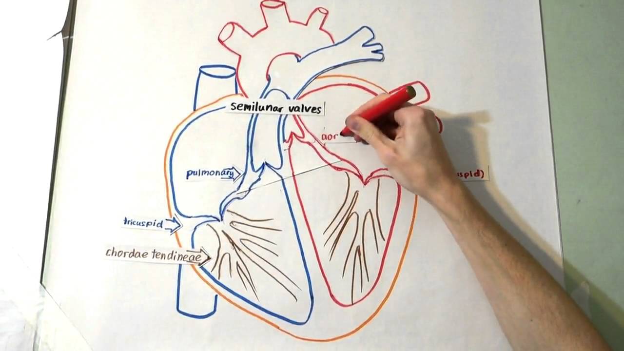
How to Draw the Internal Structure of the Heart (with Pictures) To finish drawing the aorta, draw three nubs at the top of the loop. After you draw these, erase the lines connecting from one side of the bottom of the nub to the other. Add tilted circles to the top of all of the nubs. Draw a circle at the bottom of the aorta, adjacent to the left ventricle.

Structure of the heart without labels
Anatomy of a Human Heart - uofmhealth Located between the lungs in the middle of the chest, the heart pumps blood through the network of arteries and veins known as the cardiovascular system. It pushes blood to the body's organs, tissues and cells. Blood delivers oxygen and nutrients to every cell and removes the carbon dioxide and other waste products made by those cells. Human Heart: Anatomy of Human Heart (With Diagram) | Biology ADVERTISEMENTS: In this article we will discuss about the anatomy of human heart with the help of suitable diagrams. The human heart is roughly heart-shaped structure and rests obliquely in the thoracic cavity (Fig. 7.3). The anterior surface of the human heart faces the sternum, the posterior surface—the base of the cone faces the ... Labelling the heart — Science Learning Hub Labelling the heart — Science Learning Hub Labelling the heart Add to collection The heart is a muscular organ that pumps blood through the blood vessels of the circulatory system. Blood transports oxygen and nutrients to the body. It is also involved in the removal of metabolic wastes. Topics Concepts Citizen science Teacher PLD Glossary Sign in
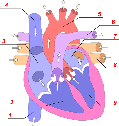
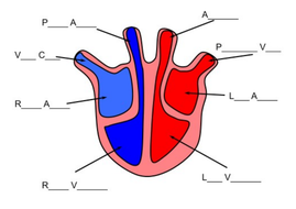
Structure of the heart without labels. Human Heart Diagram Labeled - Science Trends The heart's atrioventricular valves are structures that join the atria and ventricles of the heart together. This group of valves is comprised of the tricuspid valve and the mitral valve. Beyond this, there is a structure referred to as the aortic valve which separates the left ventricle and the aorta. Label the heart — Science Learning Hub Label the heart — Science Learning Hub Label the heart Add to collection In this interactive, you can label parts of the human heart. Drag and drop the text labels onto the boxes next to the diagram. Selecting or hovering over a box will highlight each area in the diagram. Pulmonary vein Right atrium Semilunar valve Left ventricle Vena cava The Heart Without Labels at Anatomy The heart is a muscular organ about the size of a fist, located just behind and slightly left of the breastbone. Confirm correct labels and correct misunderstandings. Region of the heart that pumps oxygenated blood to the body. This cycle is then repeated. The heart pumps blood through the network of arteries and veins called the. Heart Diagram with Labels and Detailed Explanation The heart is located under the ribcage, between the lungs and above the diaphragm. It weighs about 10.5 ounces and is cone shaped in structure. It consists of the following parts: Heart Detailed Diagram Heart - Chambers There are four chambers of the heart . The upper two chambers are the auricles and the lower two are called ventricles.
The Human Heart Without Labels at Anatomy The Human Heart Without Labels. Description this diagram shows the way blood flows through the heart. The areas of the heart with more oxygen are labeled with an "r". Heart Diagram Unlabeled Cliparts.co from cliparts.co. The heart, though small in size, performs highly significant functions that sustains human life. Heart Anatomy: Heart Dissection The major vessels of the heart are found at the base of the heart, along with the upper chambers, the right atrium (C) and left atrium (D). The atria are collapsed, but in a functioning heart, they would be stretched full of blood. The majority of the heart tissue consists of the ventricles. The left ventricle (F) is stiff and solid because it ... Heart Diagram Unlabeled - Cliparts.co 84 images of Heart Diagram Unlabeled. You can use these free cliparts for your documents, web sites, art projects or presentations. Don't forget to link to this page for attribution! Heart without labels - SlideShare 1 of 9 Heart without labels Apr. 08, 2011 • 7 likes • 7,835 views Download Now Download to read offline Education anatomy of heart from bsn class, but now modified without the labels so you can practice. John Martin Follow Lab Assistant
Structure of the Heart | The Franklin Institute Structure of the Heart Although most people know that the human heart doesn't bear much resemblance to a heart drawn on a Valentine's Day card, the image can still be a useful way to learn and remember the parts of the heart. The heart consists of four chambers: two atria on the top and two ventricles on the bottom. Heart Wall Anatomy | Structure of the Heart Wall The epicardium layer of the heart wall. 1. 2. The epicardium is a serous membrane that consists of an external layer of simple squamous and an inner layer of areolar tissue ( loose connective tissue ). The squamous cells secrete lubricating fluids into the pericardial cavity. The thick middle layer of the heart wall is called the myocardium. Structure of the Heart | SEER Training The human heart is a four-chambered muscular organ, shaped and sized roughly like a man's closed fist with two-thirds of the mass to the left of midline. The heart is enclosed in a pericardial sac that is lined with the parietal layers of a serous membrane. The visceral layer of the serous membrane forms the epicardium. Layers of the Heart Wall Diagram of Human Heart and Blood Circulation in It Four Chambers of the Heart and Blood Circulation The shape of the human heart is like an upside-down pear, weighing between 7-15 ounces, and is little larger than the size of the fist. It is located between the lungs, in the middle of the chest, behind and slightly to the left of the breast bone.
PLEASE CAN ANYONE GIVE ME A SYSTEMATIC DIAGRAM OF TH HEART - Science - - 9821775 | Meritnation.com
Structure of Heart (With Diagram) - Biology Discussion Each side of the heart consists of two chambers: (1) An upper thin walled atrium or auricle and (2) The lower thick walled chamber called as ventricle. There are two auricles or atrium right and left and two ventricles. The auricles act as receiving chambers and the ventricles the pumping chambers.
Free Blank Heart Diagram, Download Free Blank Heart Diagram png images, Free ClipArts on Clipart ...
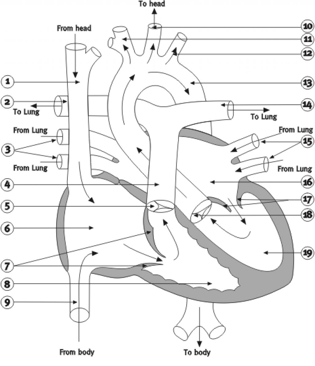
Heart Anatomy: Labeled Diagram, Structures, Function, and Blood Flow Chambers of the Heart Let's begin with the chambers of the heart. There are 4 chambers, labeled 1-4 on the diagram below. To help simplify things, we can convert the heart into a square. We will then divide that square into 4 different boxes which will represent the 4 chambers of the heart.

Easiest way to remember to draw a Human heart diagram - YouTube
Human Heart Diagram Without Labels - Labelling Worksheet This fab labelling worksheet is a great way to help your kids with their learning of the basic human anatomy. It comes with a handy heart diagram without labels, so your students can label it themselves and familiarise themselves with the different parts of the human heart. This is a really useful way of complementing your teaching of the topic ...
Structure and Function of the Heart
Heart: Anatomy and Function - Cleveland Clinic Heart. Your heart is the main organ of your cardiovascular system, a network of blood vessels that pumps blood throughout your body. It also works with other body systems to control your heart rate and blood pressure. Your family history, personal health history and lifestyle all affect how well your heart works. Appointments 800.659.7822.
34 Label Of Heart Diagram - Labels Database 2020
Human Heart - Diagram and Anatomy of the Heart - Innerbody The heart is a muscular organ about the size of a closed fist that functions as the body's circulatory pump. It takes in deoxygenated blood through the veins and delivers it to the lungs for oxygenation before pumping it into the various arteries (which provide oxygen and nutrients to body tissues by transporting the blood throughout the body).
Free Blank Heart Diagram, Download Free Blank Heart Diagram png images, Free ClipArts on Clipart ...
Human Heart (Anatomy): Diagram, Function, Chambers, Location in Body The heart is a muscular organ about the size of a fist, located just behind and slightly left of the breastbone. The heart pumps blood through the network of arteries and veins called the...
Free Heart Diagram Unlabeled, Download Free Heart Diagram Unlabeled png images, Free ClipArts on ...
Human Heart Diagram Without Labels | Human heart diagram, Heart diagram ... Human Heart Diagram Without Labels. Susan Wells. 26 followers. Human Heart Diagram. Tricuspid Valve. Carotid Artery. Heart Anatomy. Anatomy And Physiology. Biology ... Structure of the Heart. Divisions of the Heart. Circulation and the Heart. Location of the Heart. The Pericardium. Special Features of the Myocardium.

33 Label The Following Diagram Of The Heart - Labels Database 2020
A Labeled Diagram of the Human Heart You Really Need to See The human heart, comprises four chambers: right atrium, left atrium, right ventricle and left ventricle. The two upper chambers are called the left and the right atria, and the two lower chambers are known as the left and the right ventricles. The two atria and ventricles are separated from each other by a muscle wall called 'septum'.
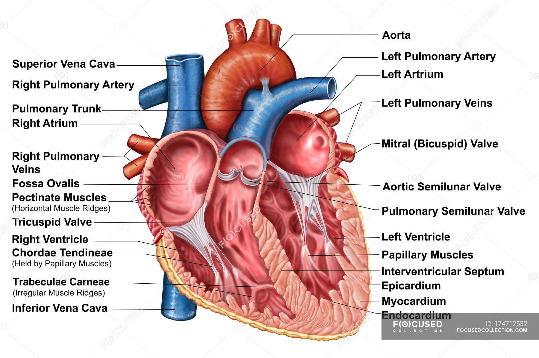
Anatomy of heart interior with labels — cross section, blood vessels - Stock Photo | #174712532
The Anatomy of the Heart, Its Structures, and Functions The heart is the organ that helps supply blood and oxygen to all parts of the body. It is divided by a partition (or septum) into two halves. The halves are, in turn, divided into four chambers. The heart is situated within the chest cavity and surrounded by a fluid-filled sac called the pericardium. This amazing muscle produces electrical ...
The Heart: Structure and Function - YouTube
The structure of the heart - Structure and function of the heart ... It is located in the middle of the chest and slightly towards the left. The heart is a large muscular pump and is divided into two halves - the right-hand side and the left-hand side. The...

Heart structure and function — structure and function of the heart cardiac output
Labelling the heart — Science Learning Hub Labelling the heart — Science Learning Hub Labelling the heart Add to collection The heart is a muscular organ that pumps blood through the blood vessels of the circulatory system. Blood transports oxygen and nutrients to the body. It is also involved in the removal of metabolic wastes. Topics Concepts Citizen science Teacher PLD Glossary Sign in
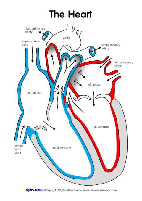
Label the Heart Worksheets (SB6634) - SparkleBox
Human Heart: Anatomy of Human Heart (With Diagram) | Biology ADVERTISEMENTS: In this article we will discuss about the anatomy of human heart with the help of suitable diagrams. The human heart is roughly heart-shaped structure and rests obliquely in the thoracic cavity (Fig. 7.3). The anterior surface of the human heart faces the sternum, the posterior surface—the base of the cone faces the ...

Heart Anatomy Quizzes and Flashcards – sciencemusicvideos
Anatomy of a Human Heart - uofmhealth Located between the lungs in the middle of the chest, the heart pumps blood through the network of arteries and veins known as the cardiovascular system. It pushes blood to the body's organs, tissues and cells. Blood delivers oxygen and nutrients to every cell and removes the carbon dioxide and other waste products made by those cells.
The Heart - AQA (9-1) | Teaching Resources

34 Label Of Heart Diagram - Labels Database 2020

Loretta Hagen Sundown Till Dawn by Richard Cuccaro Original photo: Tracy Stoft Collage w/ sky ...
Post a Comment for "40 structure of the heart without labels"